327 Central Park West (Suite 1A), New York, NY 10025

Orthodontists use “x-rays” to evaluate tooth position within the jaws and to evaluate the position of the jaws relative to each other. We often use the term radiograph when referring to an “x-ray” image.
General dentists use radiographs to visualize cavities (tooth decay) on areas of the teeth that aren’t easy to directly observe. Radiographs used by orthodontists are different than those used by general dentists because orthodontists must evaluate development and position of teeth within the bones. Orthodontists also use radiographs to look at the position of the jaws relative to the other facial bones. Radiographic information is crucial for appropriate orthodontic diagnosis and treatment.
Two types of radiographs are commonly used to generate an orthodontic diagnosis and to guide treatment – a panoramic or “pan” radiograph and a lateral cephalometric or “ceph” radiograph. The pan is obtained with a device that moves in an arc around face from ear to ear in a manner similar to the way a smart  phone is moved to create a panoramic photograph. Poorly positioned, missing or extra teeth are easy to spot in the panoramic radiograph. The shape and position of developing teeth can also be seen. We use visual information from the pan when making important decisions about treatment options that affect facial esthetics as well as the way the teeth will fit together. Asymmetric or disproportionate development of the jaws and the jaw joint can also be assessed. Pan radiographs are generally taken prior to orthodontic treatment, one year into treatment, and at the end of treatment. The pan taken one year into treatment is used to evaluate the position of the tooth roots and to assess whether the roots have shortened due to “root resorption.” If root resorption is detected, orthodontic treatment is sometimes delayed for a few months or discontinued early to avoid excessive resorption.
Prior to orthodontic treatment we also take a cephalometric radiograph. The ceph image depicts the patient 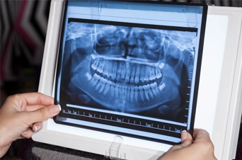 in profile (from the side of the face). The ceph allows us to visualize the front-to-back relationship of the teeth and jaws as well as the spatial relationship between the jaws and the other bones of the face. A second ceph radiograph is sometimes taken near the end of treatment. Radiation exposure from a pan or ceph radiograph is less than that received by a passenger on a typical airline flight. Nonetheless, we make every effort to reduce radiation exposure by taking the fewest necessary radiographs and by utilizing an ultra-modern digital radiograph system.
Resources:
- American Dental Association Council on Scientific Affairs. The use of dental radiographs: Update and recommendations. J Am Dent Assoc 2006;137(9):1304-12
Articles from Imagegently.org
- goo.gl/WnzRBb
- https://goo.gl/xdaJlp
[Note: All images contained in this article are courtesy of Google Images]